Welcome to AZMedicalScience — a dedicated platform for healthcare professionals to access specialized scientific content and research.

Header Logo
  • Respiratory & Immunology
  • Cardiovascular, Renal & Metabolism
  • Oncology
  • Rare Disease

Welcome to AZMedical Science

AZMedical Science is a website developed by AstraZeneca to provide non-promotional, disease state education for healthcare professionals.


Please note that the content available on this website may vary depending on your location, in accordance with local regulatory requirements.


By clicking "Enter," you confirm that you are a licensed healthcare professional and understand the nature of this content.

I am an AstraZeneca authorized person

Exit

Hypereosinophilic Syndrome (HES)

AstraZeneca Medical's ambition is to address unmet needs for people living with inflammatory diseases driven by eosinophilic inflammation.

  1. HES and Eosinophils
  2. Diagnosis and Clinical Presentation
  3. Treatment Goals and Unmet Need in HES
  4. Clinical & Economic Burden

What is HES

HES is a rare, heterogeneous condition characterized by persistently high blood eosinophil counts (≥1.5 × 10⁹ cells/L and/or tissue eosinophilia) that can lead to organ damage and/or organ dysfunction.1-3 HES can affect skin, lung, gastrointestinal, cardiac, neurologic and hematologic systems.3-5


The true incidence and prevalence of HES is unknown.6 However, data from the US and UK report an annual incidence of <1 case per 100,000 individuals.7-9

One of the most used HES definitions is the one by the ICOG-EO10

ICOG-EO consensus definition³

  • AEC ≥1.5 x 10⁹ cells/L on two examinations at least 2 weeks apart and/or tissue HEa
  • Organ damage and/or dysfunction attributable to tissue HEᵃ and exclusion of other conditions as a major reason for organ damage
  • Includes multiple clinical classifications

aHE-related organ damage (damage attributable to HE): organ dysfunction with marked tissue eosinophil infiltrates or/and extensive deposition of eosinophil-derived proteins such as eMBP1 or EPX (in the presence or absence of marked tissue eosinophils) and typical clinical, histopathological and laboratory-based signs of HE-induced organ damage.

Classifications and Subtypes of HES

HES is classified into six clinical subtypes.3,10-12

M-HES

  • Myeloproliferative HES (M-HES) with documented or presumed clonal eosinophilic involvement
  • Includes PDGFRA-associated MPN, CEL-NOS, and idiopathic HES with myeloproliferative featuresa

10–20% of patients3

Reactive HES

  • Underlying condition where eosinophils are considered non-clonal cells, and HES is considered to be cytokine driven and end-organ damage attributable to HES
  • Special variants of reactive HES include L-HES: abnormal T cells are often detected and HES-related organ damage is found

10–20% of patients3

Overlap HESb

  • Refers to single-organ eosinophilic disease and recognized multi-system eosinophilic syndromes, accompanied by peripheral eosinophilia >1.5 x 10⁹ cells/L
  • Includes eosinophilic gastrointestinal disease, single-organ chronic eosinophilic pneumonia and multi-organ EGPA

Associated HESb

  • Eosinophilia >1.5 x 10⁹ cells/L in the setting of a distinct diagnosis, in which eosinophilia has been described in a subset of affected patients
  • Includes primary immunodeficiency syndromes, such as autoimmune lymphoproliferative disease and hyper-IgE syndrome

Familial HESb

  • HES occurring in multiple members of a single family
  • Includes autosomal dominant familial HE

Idiopathic HES

  • HES of unknown cause that does not meet criteria for any of the other subtypes
  • Features multisystem involvement
  • >50% of patients3

aSuch as dysplastic eosinophils, circulating myeloid precursors, anemia, thrombocytopenia, splenomegaly, elevated serum B12, and/or tryptase levels, atypical mast cells, hypercellular marrow, and myelofibrosis.

bThe relative frequencies of these variants in the general population are unclear, due in part to the lack of universally accepted definitions of HES and referral bias.3

eosinophills-new.png

What are Eosinophils?

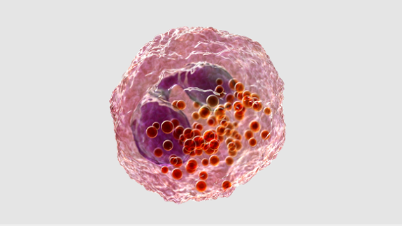
Eosinophils are terminally differentiated granulocytes that play key roles in host defense, tissue remodeling and inflammation. They release cytotoxic granule proteins (EDN, ECP, EPO, MBP), extracellular traps, and lipid body inflammatory mediators and interact with other immune cells to perpetuate inflammation.13-15

Eosinophil overproduction may arise from clonal expansion, cytokine dysregulation or other unknown mechanisms

Clonal eosinophilic proliferation16

  • A primary molecular defect involving hematopoietic stem cells and/or
  • Defects in signal transduction from the receptors that mediate eosinophilopoiesis.

Overproduction of eosinophilopoietic cytokines, such as IL-513,16

  • Involvement of cytokine overproduction and IL-5 in eosinophil production.

Other mechanisms3,16

  • Defects in normal suppressive regulation of eosinophil survival and activation
  • Abnormalities in chemotaxis
  • Functional abnormalities of eosinophilopoietic cytokines related to enhanced or prolonged biologic activity (though not conclusively demonstrated).

Tissue infiltration

Overproduction and tissue infiltration of eosinophils leads to organ damage in HES.17

Tissue Infilteration

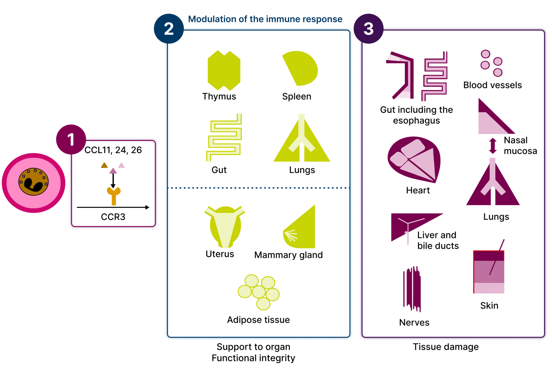
An array of chemokines targeting the chemokine receptor CCR3 promote eosinophil recruitment into organs and tissues.1

A first set of target tissues hosts a population of regulatory eosinophils involved in the maintenance of immune homeostasis and functional integrity of specific organs.1

Other tissues are targets for eosinophil infiltration during inflammation – commonly the gut, lungs and skin.¹

AECAbsolute Eosinophil Count
CCLChemokine Ligand
CCRChemokine Receptor
CEL-NOSChronic Eosinophilic Leukemia-Not Otherwise Specified
ECPEosinophil Cationic Protein
EDNEosinophil-Derived Neurotoxin
EGPAEosinophilic Granulomatosis with Polyangiitis
eMBP1Eosinophil Major Basic Protein 1
EPOEosinophil Peroxidase
EPXEosinophil Peroxidase
HEHypereosinophilia
ICOG-EOInternational Cooperative Working Group on Eosinophil Disorders
IgEImmunoglobulin E
ILInterleukin
L-HESLymphocytic Hypereosinophilic Syndrome
MBPMajor Basic Protein
MPNMyeloproliferative Neoplasm
PDGFRAPlatelet-Derived Growth Factor Receptor Alpha
UKUnited Kingdom
USUnited States

1. Chen MM, et al. J Allergy Clin Immunol Pract. 2022;10(5):1217‒1228.e3.


2. Gotlib J. Am J Hematol. 2017;92(11):1243‒1259.


3. Valent P, et al. Allergy. 2023;78(1):47–59.


4. Wechsler ME, et al. J Allergy Clin Immunol. 2023;151(6):1415–1428.


5. Ogbogu PU, et al. J Allergy Clin Immunol. 2009;124(6):1319–1325.e3.


6. Mikhail ES, Ghatol A. Hypereosinophilic Syndrome. StatPearls Publishing; 2024. Updated 11 January 2024. Accessed 12 February 2026. www.ncbi.nlm.nih.gov/books/NBK599558


7. Nguyen L, et al. Cancers. 2024;16(7):1383.


8. Shomali W, Gotlib J. Am J Hematol. 2022;97(1):129–148.


9. Requena G, et al. Immun Inflamm Dis. 2021;9(4):1447–1451.


10. Klion AD. Hematology Am Soc Hematol Educ Program. 2022;2022(1):47–54.


11. Klion AD. Hematology Am Soc Hematol Educ Program. 2015;2015:92–97.


12. Klion AD. Blood. 2015;126(9):1069–1077.


13. Ackerman SJ, Bochner BS. Immunol Allergy Clin North Am. 2007;27(3):357–375.


14. Jacobsen EA, et al. Annu Rev Immunol. 2021;39:719–757.


15. Cogan E, Roufosse F. Expert Rev Hematol. 2012;5(3):275–290.


16. Leru PM. Clin Transl Allergy. 2019;9:36.


17. Ramirez GA, et al. BioMed Res Int. 2018;2018:9095275.

Diagnosis of HES Is a Complex Clinical Challenge

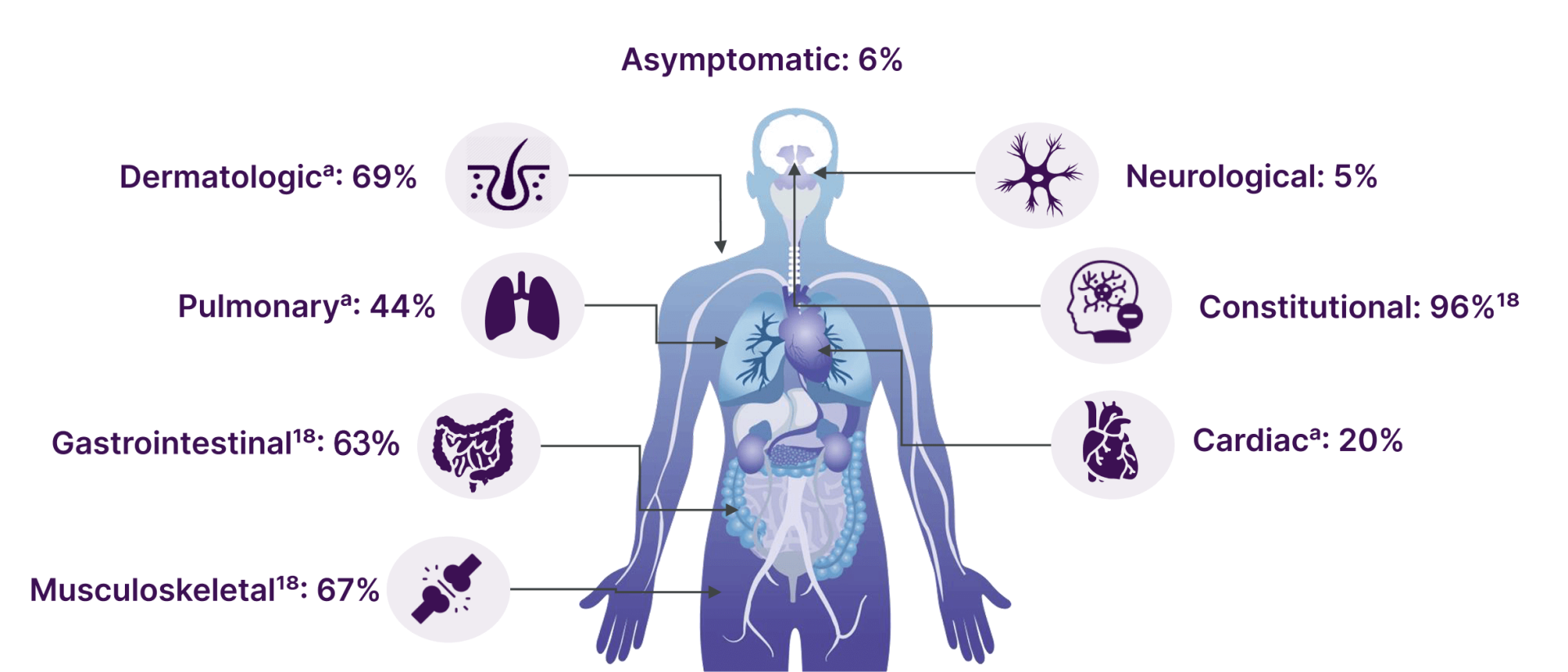
HES can manifest as tissue-specific or widespread organ damage, resulting in diverse symptoms.1

Diagnosis of HES

Some patients had multiple simultaneous manifestations. Percentages can vary depending on center and referral bias related to physician profile and sub-specialty.1


aPercentages represent the proportion of patients with subsequent clinical manifestation at the time of retrospective analysis.

Data from a real-world retrospective study suggest that >50% of patients with L-HES and I-HES at baseline and >60% of patients at year 1 of follow-up have ≥3 different organ systems with HES signs and/or symptoms.2

Common HES Manifestations

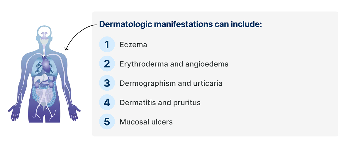
Dermatologic manifestations

Pulmonary manifestations

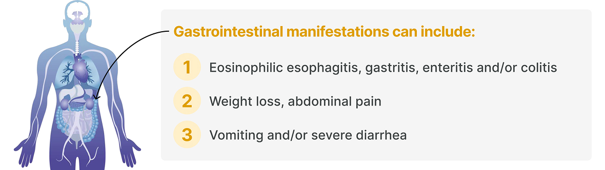
Gastrointestinal manifestations

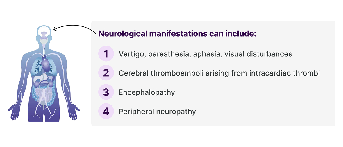
Neurological Manifestations

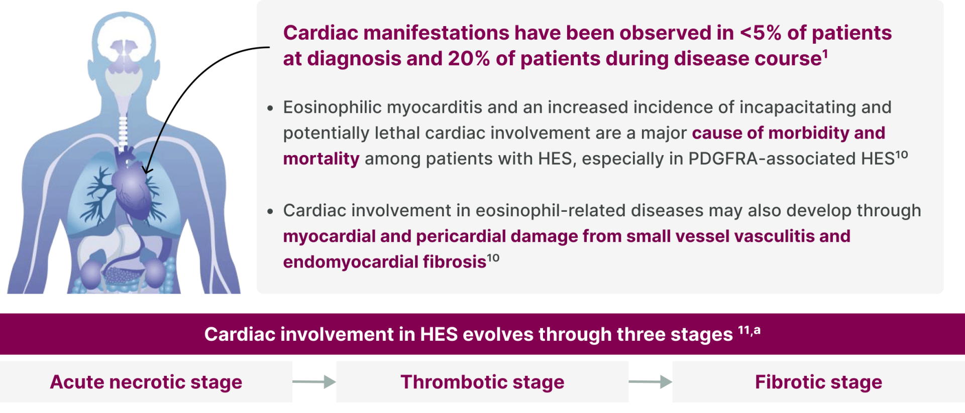
Cardiac manifestations

ᵃCardiac complications in HES typically develop in a stepwise manner, although overlap between successive phases may occur.

Diagnostic Challenges

HES is characterized by diagnostic delays and is often misunderstood and may be misdiagnosed because of its varied presentation, which can lead to patients seeing multiple specialists before their condition is recognized.12


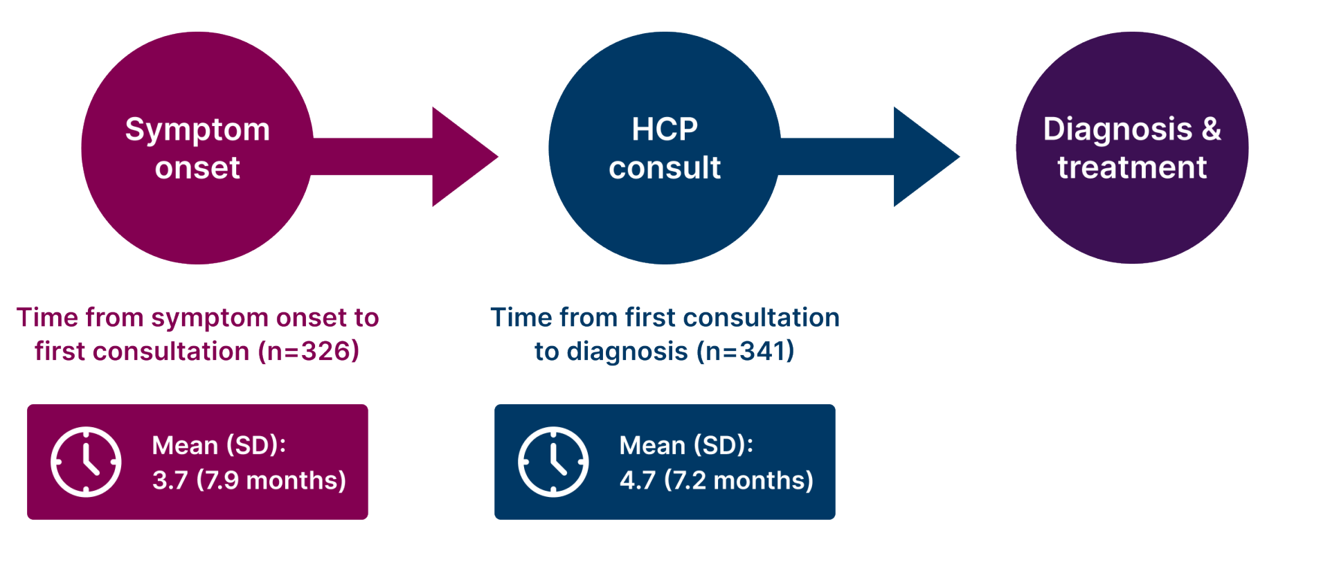
Often months pass between symptom onset to first consultation and toward diagnosis of HES.13,a

Tissue Infilteration

aHCP types that were consulted by patients included PCP/GP, hematologist, allergist/immunologist, dermatologist, gastroenterologist, rheumatologist, pulmonologist and internist, cardiologist, and neurologist.


Considerations for Diagnosis

Hypereosinophilia is a defining feature of HES14

Multiple potential causes of eosinophilia must be ruled out before diagnosis of HES, including15:

Infections

Medications

Hematologic/neoplastic disorders

Immune dysregulation

Allergic disorders

Other

A. Physical examination and detailed history16

Assess for signs associated with clonal (neoplastic) HE14,16:


  • Risk factors and family history of cancera


Screen for secondary (reactive) causes of HE16,17:


  • Evidence of primary immunodeficiencyb
  • Drug exposure (including non-prescription)
  • Travel history and lifestyle associated with risk of exposure to helminthic parasites

B. Microscopic examination of blood and body fluids16

Test for infections which cause secondary (reactive) HE16,17:


  • Ova/larvae or helminthic parasites (such as Toxocara, Strongyloides, Ascaris)

C. Blood test/bone marrow exam16,c

Seek evidence for HES including secondary (reactive) causes of HE16:


  • CBC with differential absolute eosinophil count and peripheral blood smear
  • Serum tryptase, vitamin B12, IgE, IgG, IgM and IgG4
  • ANCAd
  • T-cell immunophenotyping by flow cytometry
  • Assessment for and quantification of blasts
  • Cytogenetic and molecular testing for PDGFRA/B, FGFR1, JAK2 rearrangements or mutations associated with clonal eosinophilia, TCR gene rearrangement analysis by PCR/NGS

aClonal (neoplastic) HE is associated with underlying stem cell, myeloid, or eosinophil neoplasm, inducing HE14; 


bHE may be associated with primary immunodeficiency disorders17; 


cBone marrow examination should be considered in the following situations: phenotyping and/or cytogenetic tests suggesting myeloid or lymphoid variant HES, blood eosinophilia greater than 5 x 10⁹ cells/L, elevated serum tryptase and lack of response to systemic corticosteroid treatment16;

dAnti-PR3/anti-MPO testing by ELISA if ANCA detected.16

Cardiac

  • Serum troponin
  • Echocardiogram electrocardiogram
  • Cardiac MRI
  • Endomyocardial biopsy

Hematologic

  • Bone marrow biopsy
  • CT scans of chest/abdomen/pelvis 
  • FDG-PET scan
  • Peripheral blood FISH/RT-PCR

Dermatologic

  • Skin biopsy

Gastrointestinal

  • GI endoscopy with biopsies
  • Abdominal CT scan
  • Blood chemistry: liver enzymes, amylase/lipase
  • Liver biopsy

Neurologic

  • Brain MRI or CT with contrast
  • Electroencephalogram
  • Nerve conduction studies

Vascular

  • Venous doppler ultrasound
  • Angiography

Renal

  • Kidney biopsy
  • Urinalysis

Pulmonary

  • PFT
  • Bronchoscopy with bronchoalveolar lavage and/or lung biopsy
  • Chest X-ray and/or CT

ANCAAnti-Neutrophil Cytoplasmic Antibodies
CBCComplete Blood Count
CTComputed Tomography
ELISAEnzyme-Linked Immunosorbent Assay
FDG-PETFluorodeoxyglucose Positron Emission Tomography
FGFR1Fibroblast Growth Factor Receptor 1
FISHFluorescence In Situ Hybridization
GIGastrointestinal
GPGeneral Practitioner
HCPHealth Care Provider
HEHypereosinophilia
IgImmunoglobulin
I-HESIdiopathic Hypereosinophilic Syndrome
JAK2Janus Kinase 2
L-HESLymphocytic Hypereosinophilic Syndrome
MPOMyeloperoxidase
MRIMagnetic Resonance Imaging
NGSNext Generation Sequencing
PCPPrimary Care Provider
PCRPolymerase Chain Reaction
PDGFRA/BPlatelet-Derived Growth Factor Receptor Alpha/Beta
PFTPulmonary Function Test
PR3Proteinase 3
RT-PCRReverse Transcription-Polymerase Chain Reaction
SDStandard Deviation
TCRT-Cell Receptor

1. Ogbogu PU, et al. J Allergy Clin Immunol. 2009;124(6):1319–1325.e3.


2. Dolin P, et al. Poster presented at: ASH; December 7-10, 2024; San Diego, CA. Poster P2316.


3. Leiferman KM, Peters MS. J Allergy Clin Immunol Pract. 2018;6(5):1462–1482.e6.


4. Kovacs N, et al. J Allergy Clin Immunol Pract. 2020;8(9):3209–3212.e8.


5. Dulohery MM, et al. Respir Med. 2011;105(1):114–121.


6. Zhang L, et al. Pulm Circ. 2018;9(1):1–4.


7. Kuang FL, et al. J Allergy Clin Immunol Pract. 2020;8(8):2718–2726.e2.


8. Parfitt JR, et al. Mod Pathol. 2006;19(1):90–96.


9. Jonakowski M, et al. Mult Scler Relat Disord. 2021;51:102871.


10. Ogbogu PU, et al. Immunol Allergy Clin North Am. 2007;27(3):457–475.


11. Bondue A, et al. Heart. 2022;108(3):164–171.


12. Wechsler ME, et al. J Allergy Clin Immunol. 2023;151(6):1415–1428.


13. Dolin P, et al. Poster presented at: EHA; June 13-16, 2024; Madrid, Spain. Poster P2008.


14. Valent P, et al. Allergy. 2023;78(1):47–59.


15. Curtis C, Ogbogu P. Clin Rev Allergy Immunol. 2016;50(2):240‒251.


16. Khoury P, et al. Mayo Clin Proc. 2023;98(7):1054–1070.


17. Nguyen L, et al. Cancers. 2024;16(7):1383.


18. Silver J, et al. J Allergy Clin Immunol Glob. 2025;4(3):100501.

HES Management Considerations

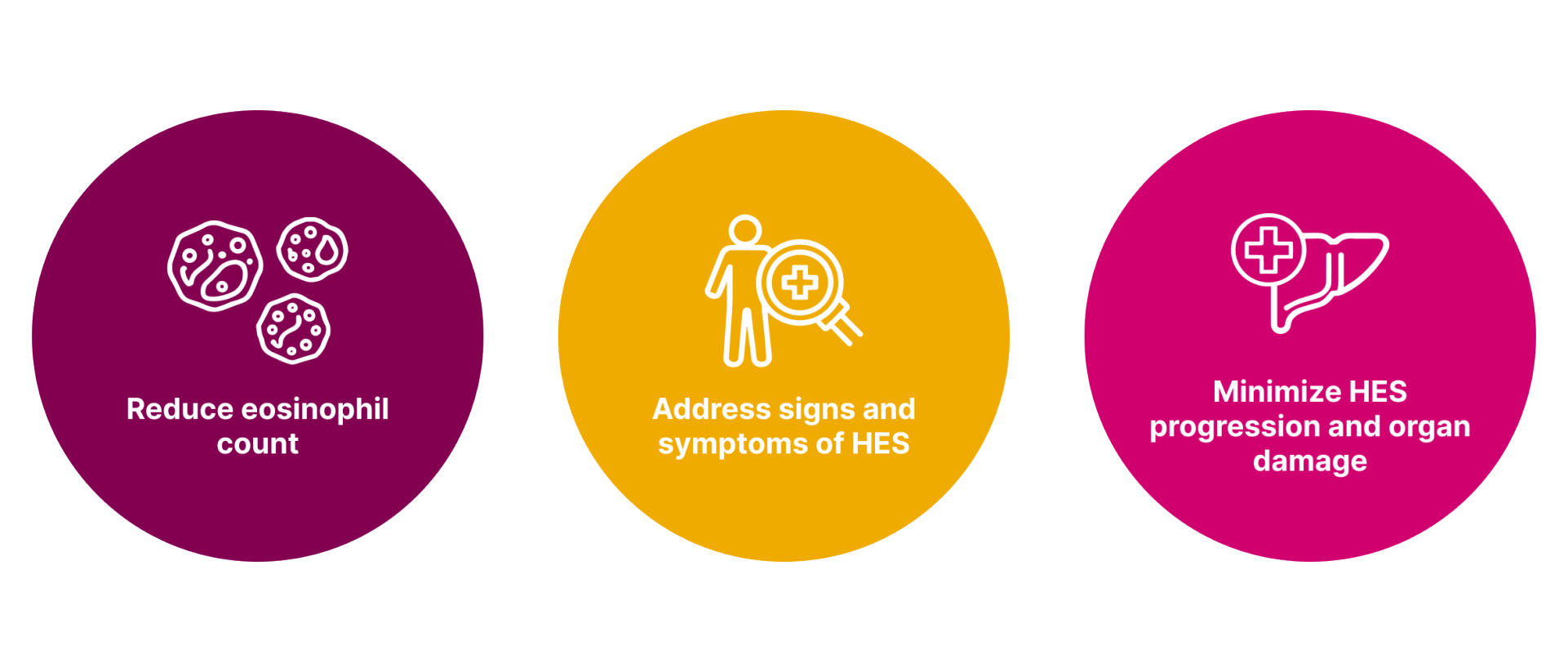
HES treatment considerations include controlling inflammation, addressing symptoms, and preventing disease relapse, while minimizing dose- and drug-related toxicity.6,7

HES Management Considerations

Decreasing eosinophil may alleviate inflammation and related symptoms, while preventing disease progression.7

Managing HES

There is a lack of comprehensive and clear guidelines for the treatment, management and follow-up of patients with HES.1,2


In most cases, glucocorticoids are the mainstay of HES therapy, although it may differ in patients with FIP1L1-PDGFRA-positive disease.1,2


However, lack of efficacy and intolerance due to treatment-related side effects can lead to discontinuation of therapy in a large proportion of patient cases.2

Treatments Utilized in HES1,2

Treatments Utilized in HES Treatments Utilized in HES

HES can have a negative impact on the quality of life of patients3

HES can impact multiple health domains. The overall physical impact can vary greatly across patients, depending on how organs are impacted. The majority of patients with HES experience negative impacts on walking ability, sleep, and feeling fatigued. Quality-of-life impacts may include the following:

Social impact

relationships with others, social activities and outings, work/school activities.

Emotional impact

feeling depressed/hopeless, anxious/worried, embarrassed/ashamed, cranky/irritated, or frustrated/annoyed.

Global physical impact

negative impact on walking ability, sleep, and feeling fatigued more often.

Cognitive impact

difficulty concentrating/memory problems, change in behavior, difficulty having conversations, or delayed reactions.

HES can impact multiple quality of life domains

Impacts of HES Impacts of HES

Overall health status, impairment and fatigue increase as HES disease severity increases.4

HES presents a mortality risk, primarily due to cardiac dysfunction, infection, thromboembolic phenomena and vascular disease.5

FIP1L1-PDGFRAFusion of Factor Interacting with PAPOLA and PDGF Receptor Alpha

1. Wechsler ME, et al. J Allergy Clin Immunol. 2023;151(6):1415–1428.


2. Ogbogu PU, et al. J Allergy Clin Immunol. 2009;124(6):1319–1325.e3.


3. Kovacs N, et al. J Allergy Clin Immunol Pract. 2020;8(9):3209–3212.e8.


4. Schwaab J, et al. Poster presented at: ASH; December 7-10, 2024; San Diego, CA. Poster 5075.


5. Podjasek JC, Butterfield JH. Leuk Res. 2013;37(4):392–395.


6. Kuang FL, Klion AD. J Allergy Clin Immunol Pract. 2017;5(6):1502–1509.


7. Nguyen L, et al. Cancers. 2024;16(7):1383.

Clinical and Economic Burden of HES

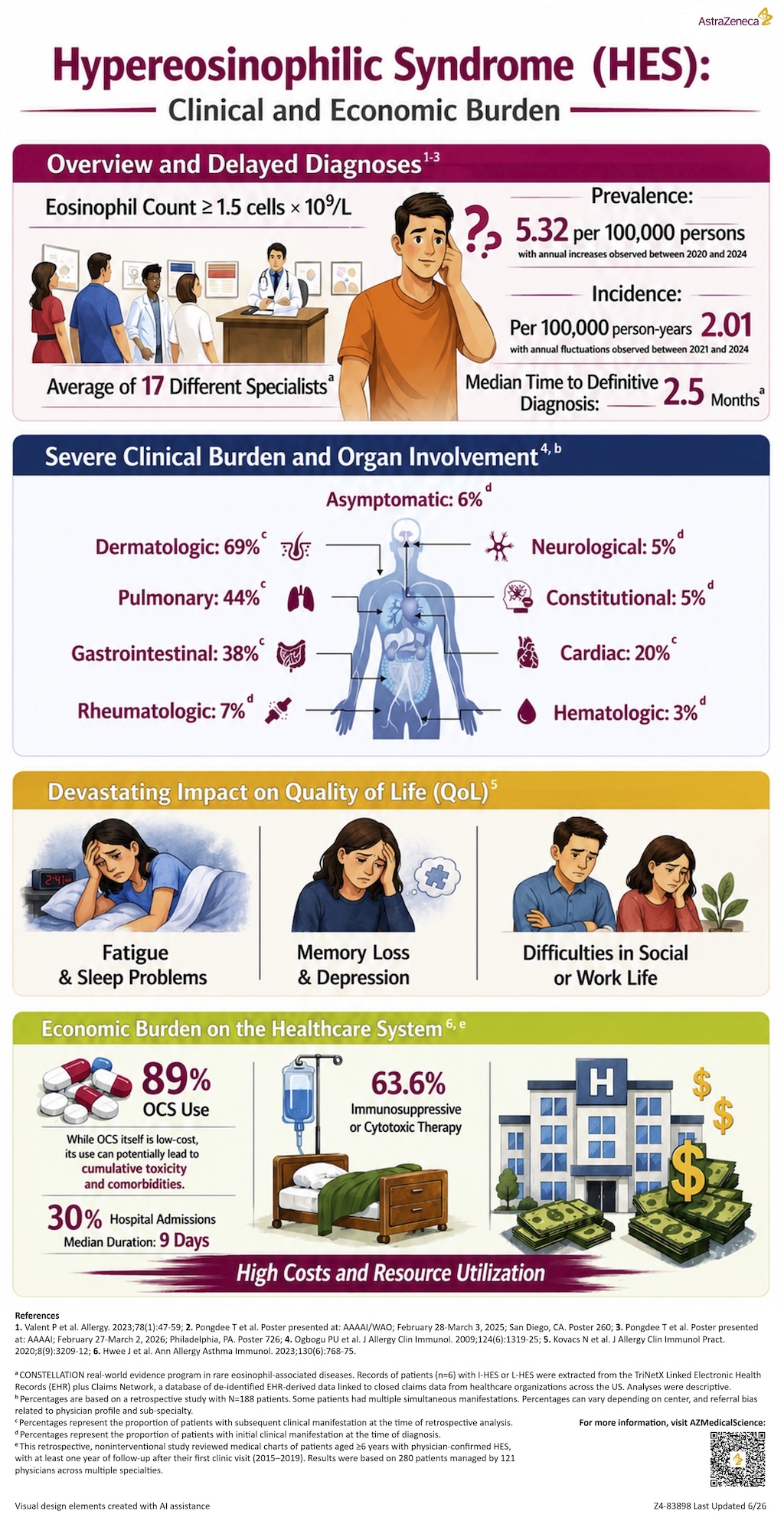
HES is a rare disorder characterized by persistent hypereosinophilia that can lead to multisystem organ damage.1-4 It is associated with significant diagnostic delays, impaired quality of life, and substantial healthcare resource utilization — underscoring its clinical and economic burden.5,6

Filename
clinical-and-economic-burden-of-hypereosinophic-syndrome.pdf
Size
3 MB
Format
PDF
Download
Hypereosinophilic syndrome (HES) infographic highlighting clinical burden, prevalence and incidence rates, delayed diagnosis, organ involvement, quality of life impact, and healthcare costs including steroid use and hospital admissions.

1. Valent P et al. Allergy. 2023;78(1):47-59.


2. Pongdee T et al. Poster presented at: AAAAI/WAO; February 28-March 3, 2025; San Diego, CA. Poster 260.


3. Pongdee T et al. Poster presented at: AAAAI; February 27-March 2, 2026; Philadelphia, PA. Poster 726.


4. Ogbogu PU et al. J Allergy Clin Immunol. 2009;124(6):1319-25.


5. Kovacs N et al. J Allergy Clin Immunol Pract. 2020;8(9):3209-12.


6. Hwee J et al. Ann Allergy Asthma Immunol. 2023;130(6):768-75.

Z4-84755 Last Updated 6/26

Connessiallasalute.it

This site is intended for healthcare professionals


ASTRAZENECA Z4-85831 | 8/2026 |  ©ASTRAZENECA 2025 ALL RIGHTS RESERVED


Cookies

Contact Us

Privacy Notice