Welcome to AZMedicalScience — a dedicated platform for healthcare professionals to access specialized scientific content and research.

Header Logo
  • Respiratory & Immunology
  • Cardiovascular, Renal & Metabolism
  • Oncology
  • Rare Disease

Welcome to AZMedical Science

AZMedical Science is a website developed by AstraZeneca to provide non-promotional, disease state education for healthcare professionals.


Please note that the content available on this website may vary depending on your location, in accordance with local regulatory requirements.


By clicking "Enter," you confirm that you are a licensed healthcare professional and understand the nature of this content.

I am an AstraZeneca authorized person

Exit

IL-33 in COPD

Explore the latest in COPD science
  • Home
  • Burden of COPD
  • Inflammation in COPD
  • Mucus dysfunction in COPD
  • IL-33 in COPD
  • Resources

Snapshot

IL-33 is highly expressed in severe COPD and may be associated with productive cough and future exacerbation risk, highlighting IL-33 as a driver of
disease activity1–3

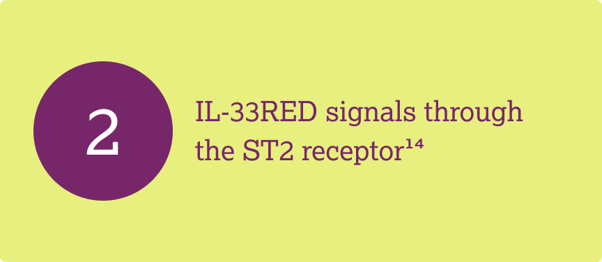
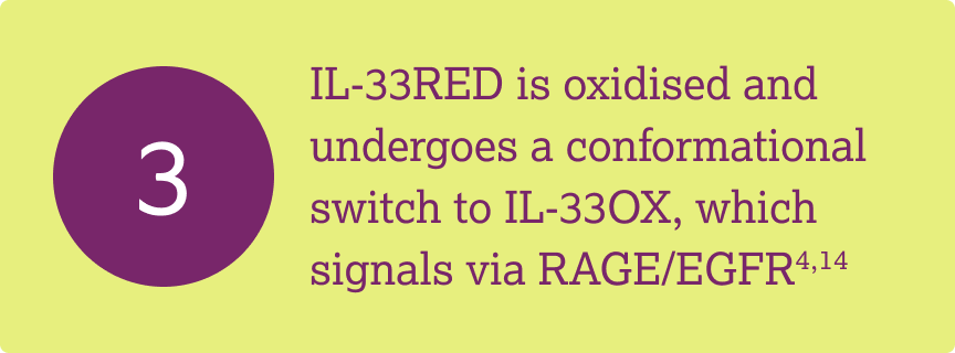
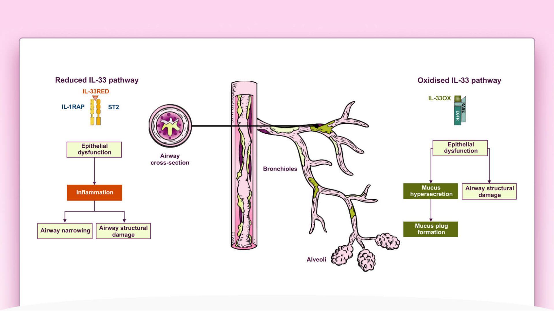
IL-33RED signals via ST2 to activate broad Type 1, Type 2 and Type 3 inflammation, while oxidized IL-33OX signals via RAGE/EGFR in epithelium to promote mucus hypersecretion
and impaired
epithelial repair4–7

Watch the role of IL-33 in COPD

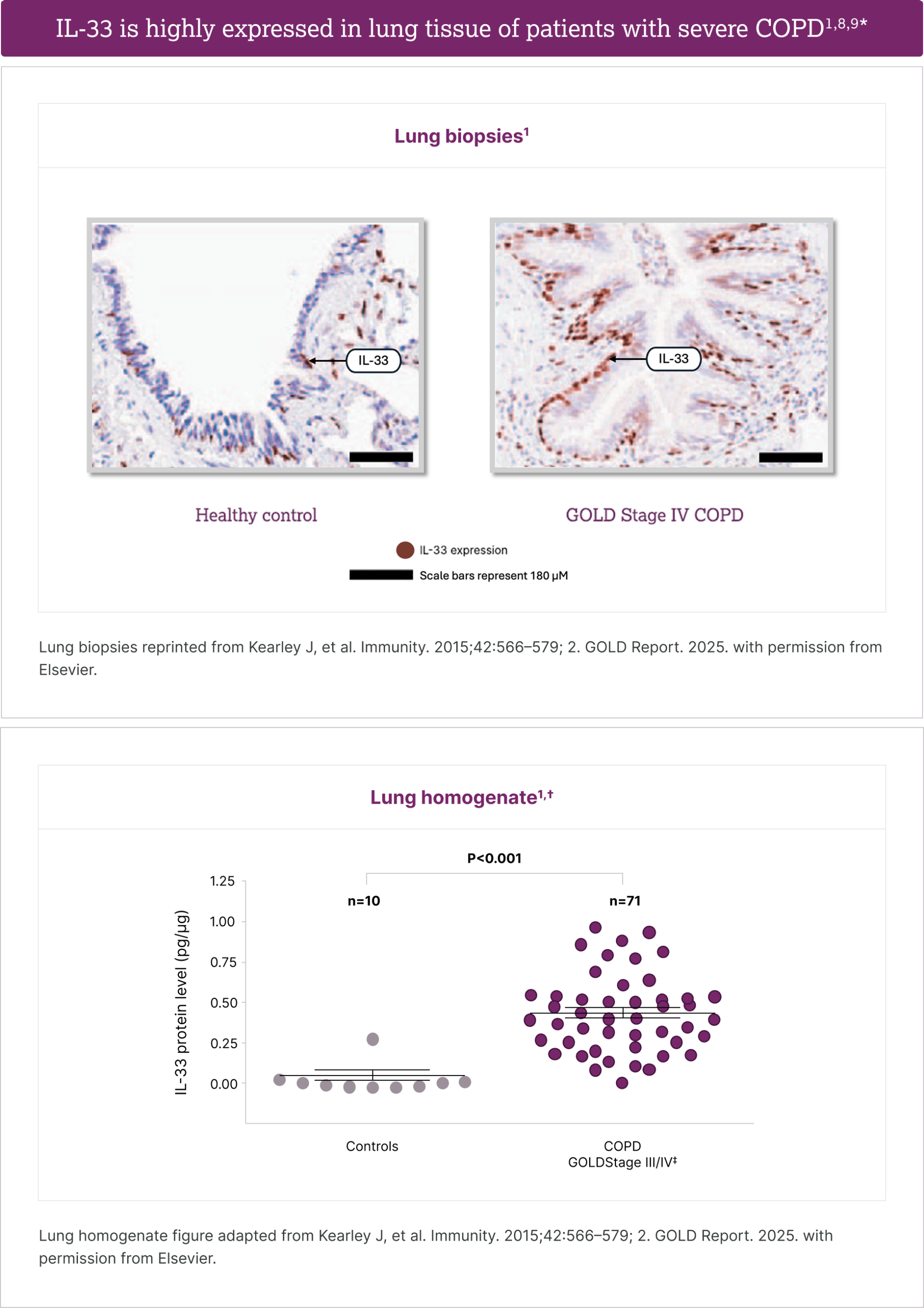
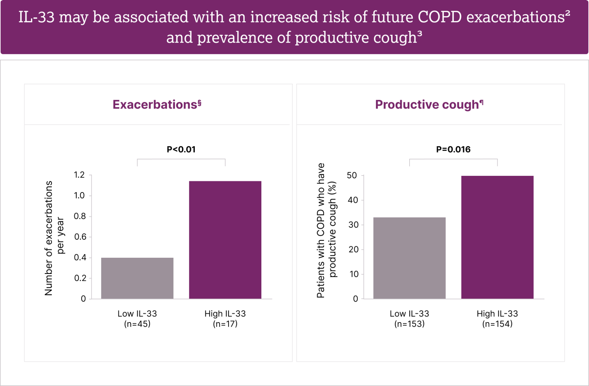
IL-33 is highly expressed in lung tissue samples from patients with severe COPD1, and may be associated with increased risk of future COPD exacerbations2 and prevalence of
productive cough3

View more

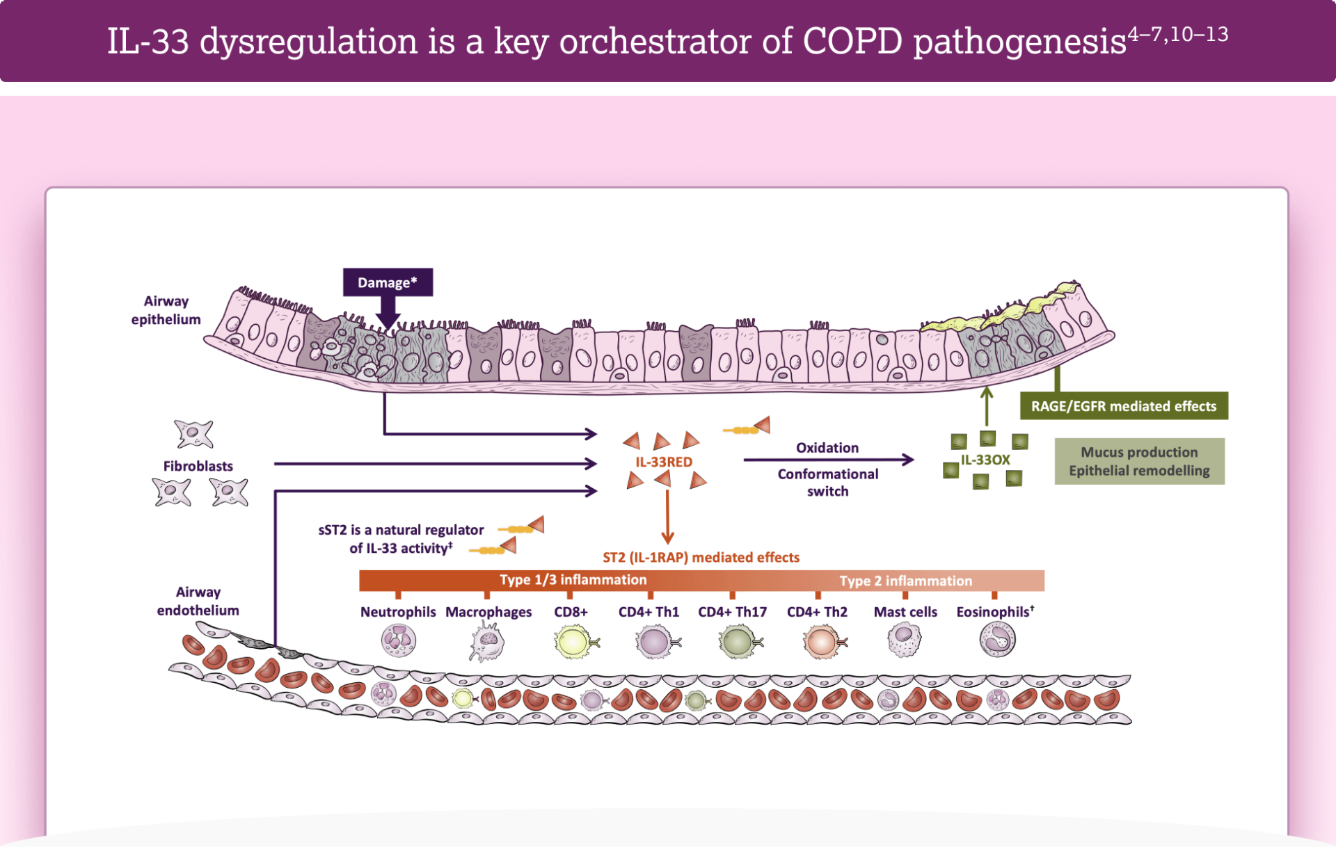
IL-33 dysregulation is a key orchestrator of

COPD pathogenesis4–7,10–13

View more

Dysregulation of the dual pathway of IL-33 may have pathological consequences4,5,18,19

View more

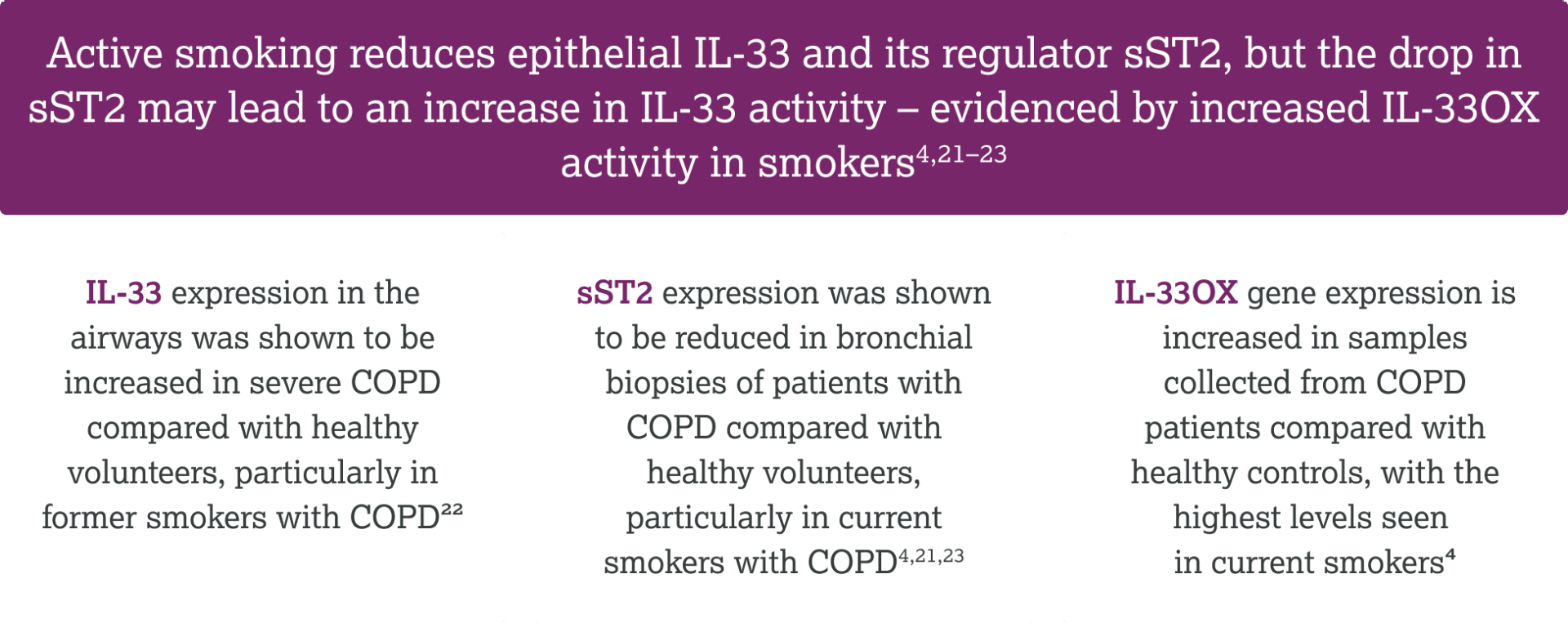
Smoking can influence expression of the
IL-33 pathway4,21,22

View more
  1. <h4>Over half of patients with COPD receiving triple inhaled therapy have been shown to continue to experience exacerbations<sup>1,2</sup></h4>
  2. <h4>Over half of patients with COPD receiving triple inhaled therapy have been shown to continue to experience exacerbations<sup>1,2</sup></h4>
  3. <h4>Over half of patients with COPD receiving triple inhaled therapy have been shown to continue to experience exacerbations<sup>1,2</sup></h4>
  4. <h4>Over half of patients with COPD receiving triple inhaled therapy have been shown to continue to experience exacerbations<sup>1,2</sup></h4>

Mucus dysfunction in COPD

Previous

Resources

Next

Frequently asked questions

What does ‘dysregulated IL-33’ mean?

IL‑33 is released upon stress or cell damage to initiate a host response—what was adaptive turns maladaptive or ‘dysregulated’ when its levels chronically surpass buffering mechanisms and signalling is persistent1–8

How does smoking affect the IL-33 pathway in patients with COPD?

In COPD, smoking status affects the IL‑33 axis—former smokers show elevated IL-33 compared to current smokers, while current smokers show reduced sST2 and more oxidized IL‑33, indicating aberrant IL‑33–ST2 signalling in both groups9–12

How does the role of IL-33 in COPD differ from other epithelial cytokines?

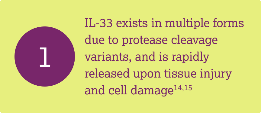
Damage to the epithelium triggers the release of upstream, alarmin cytokines. IL-33 is pre-formed, rapidly released and initiates a broad inflammatory cascade encompassing Type 1, Type 2 and Type 3 responses1,13,14

What evidence would suggest my patient with COPD has dysregulated IL-33 signalling?

Frequent exacerbations, productive cough and persistent airway/systemic inflammation may suggest heightened IL‑33

pathway activity15–17

What are the potential consequences of dysregulated IL-33 signalling?

IL-33 through its dual forms can drive Type 1 and 3 and Type 2 (eosinophilic, subset) inflammation, and mucus dysfunction, which are key drivers of COPD pathogenesis1–3,12,15

What is mucus dysfunction and what is the impact for patients with COPD?

Mucus dysfunction is the pathological changes in mucus volume, composition and clearance, the airway manifestations of these pathological changes, and the downstream

clinical consequences12,15,18–25

  1. sss
  2. sss
  3. sss
  4. sss
  5. sss
  6. sss

  1. Cayrol C. Cells. 2021;11(1):107
  2. Calderon AA, et al. Eur Respir Rev. 2023;32(167):220144
  3. Brightling CE, et al. Eur Respir J. 2019;54(6):1900651
  4. England E, et al. Sci Rep. 2023;13(1):9825
  5. Rabe KF, et al. Am J Respir Crit Care Med. 2023;208(4):395–405
  6. Keddache S, et al. Clin Immunol. 2021;229:108798
  7. Scott IC, et al. Biochem Soc Trans. 2018;46(5):1345–1353
  8. Burgoyne RA, et al. Cells. 2021;10(10):2763
  9. Abdo M, et al. Eur Respir J. 2024;64(3):2400347
  10. Faiz A, et al. Am J Respir Crit Care Med. 2023;208(10):1075–1087
  11. Faiz A, et al. Am J Respir Crit Care Med. 2023;208(Suppl.):1075–1087
  12. Strickson S, et al. Eur Respir J. 2023;62(3):2202210
  13. Cayrol C, Girard JP. Cytokine. 2022;156:155891
  14. Varricchi G, et al. Eur Respir J. 2024;63(4):2301619
  15. Diaz AA, et al. JAMA. 2023;329(21):1832–9
  16. Shen Y, et al. Int J Chron Obstruct Pulmon Dis. 2018;13:399–407
  17. Rodrigues SO, et al. Pharmaceuticals (Basel). 2021;14(10):979
  18. Fahy JV, Dickey BF. N Engl J Med. 2010;363(23):2233–2247
  19. Stott-Miller M, et al. Int J Chron Obstruct Pulmon Dis. 2020;15:2467–2476
  20. Hughes R, et al. Respir Med. 2022;200:106921
  21. Choate R, et al. Chronic Obstr Pulm Dis. 2020;7(1):49–59
  22. Cook N, et al. Int J Chron Obstruct Pulmon Dis. 2019;14:1365–1376
  23. Tian PW, Wen FQ. J Transl Int Med. 2015;3(3):89-92
  24. Mettler SK, et al. N Engl J Med. 2025;392(19):1973–1975
  25. Zhou D, et al. Expert Rev Respir Med. 2025:1–7

Stay ahead

Sign up for the latest updates on new content

Sign up

Back to top

CD4+, T helper cells; CD8+, cytotoxic T lymphocytes; COPD, chronic obstructive pulmonary disease; EGFR, epidermal growth factor receptor; GOLD, Global Initiative for Chronic Obstructive Lung Disease; GSVA, gene set variation analysis; HV, healthy volunteers; IL, interleukin; IL-1RAP, interleukin-1 receptor accessory protein; NS, not significant; OX, oxidised; RAGE, receptor for advanced glycation endproducts; RED, reduced; ST2, serum stimulation-2; sST2, soluble serum stimulation-2

  1. Kearley J, et al. Immunity. 2015;42:566–579
  2. Joo H, et al. BMC Pulm Med. 2021;21:86
  3. Kim SW, et al. Int J Chron Obstruct Pulmon Dis. 2017;12:395–402
  4. Strickson S, et al. Eur Respir J. 2023;62:2202210
  5. Calderon AA, et al. Eur Respir Rev. 2023;32:220144
  6. Cohen ES, et al. Nat Commun. 2015;6:8327
  7. Brightling C, Greening N. Eur Resp J. 2019;54:1900651
  8. GOLD Report. 2025. Available from: https://goldcopd.org/2025-gold-report/ (Accessed June 2026)
  9. Kearley J, et al. Immunity. 2015;42 (Suppl.):566–579
  10. Wechsler ME, Wells JM. ERJ Open Res. 2024;10:00177–02024
  11. Marcuccio G, et al. Front Med Lausanne. 2025;12:1550716
  12. Gabryelska A, et al. Front Immunol. 2019;10:692
  13. Zhou Y, et al. J Transl Med. 2023;21:902
  14. Cayrol C. Cells. 2021;11:107
  15. Cayrol C, Girard J-P. Cytokine. 2022;156:155891
  16. Griesenauer B, Paczesny S. Front Immunol. 2017;8:475
  17. Scott IC, et al. Biochem Soc Trans. 2018;46:1345–1353
  18. MacNee W. Br Med J. 2006;332:1202–1204
  19. Tian P-W, Wen F-Q. J Transl Int Med. 2015;3:89–92
  20. Diaz AA, et al. JAMA. 2023;329(21):1832–9
  21. Faiz A, et al. Am J Respir Crit Care Med. 2023;208:1075–1087
  22. Abdo M, et al. Eur Respir J. 2024;64:2400347
  23. Faiz A, et al. Am J Respir Crit Care Med. 2023;208(Suppl.):1075–1087

fifth-modal fifth-modal

*Damage induced by smoke, pollutants and viral or bacterial exposure7,10; †Neutrophilic inflammation is seen in the majority of patients with COPD, and up to one third of patients may also have elevated eosinophils5


Diagram developed by AstraZeneca based on information and methodology described in references4–7,10–13

modal modal
modal2 modal2
modal3 modal3
modal4 modal4

  • Dysregulated IL-33RED signalling may cause airway narrowing and airway structural damage, resulting in worsening of COPD symptoms and increased exacerbation risk4,5
  • Dysregulated IL-33OX signalling may cause mucus hypersecretion, mucus plugging and reduced airway epithelial repair, which may result in increased symptoms, risk of exacerbation and disease progression4,19,20
second-modal

Diagram developed by AstraZeneca based on information and methodology described in references4,5,20

third-modal third-modal

second-modal second-modal

*GOLD Stage III defined as severe COPD (FEV1  predicted ≥30–<50% ); GOLD Stage IV defined as very severe COPD (FEV1 predicted <30%);8 †Data are shown as individual values; horizontal lines indicate median;1 ‡This cohort comprised 17 GOLD Stage III and 54 Stage IV patients with COPD undergoing lung volume reduction surgery. Lung tissue from 10 control individuals was obtained from people who died from non-respiratory causes.9

second-modal second-modal

Exacerbations figure (left) adapted from Joo H, et al. BMC Pulm Med. 2021:21:86. Productive cough figure (right) developed using data in Kim SW, et al. Int J Chron Obstruct Pulmon Dis. 2017;12:395–401.


All figures licensed under CC BY 4.0 (https://creativecommons.org/licenses/by/4.0/).


§In this analysis of 62 patients with COPD based in Korea, levels of IL-33 in the upper quartile of the cohort were defined as high group, with all levels below this value defined as low group. Patients were prospectively followed for 1 year and monitored for exacerbation;2 ¶In this analysis of 307 patients from the COPD Korean Obstructive Lung Disease cohort, IL-33 levels above the median IL-33 level of the cohort were defined as high, with all values below the median defined as low. At baseline, the median IL-33 level was 11.9 pg/mL (IQR 7.9–30.6). This study described productive cough as chronic bronchitis, which was defined as phlegm for ≥3 months per year3

Z4-86693 | Date of preparation: September 2026

Sign up below to be notified about new resource

Connessiallasalute.it

This site is intended for healthcare professionals


ASTRAZENECA Z4-85831 | 8/2026 |  ©ASTRAZENECA 2025 ALL RIGHTS RESERVED


Cookies

Contact Us

Privacy Notice