Welcome to AZMedicalScience — a dedicated platform for healthcare professionals to access specialized scientific content and research.

Header Logo
  • Respiratory & Immunology
  • Cardiovascular, Renal & Metabolism
  • Oncology
  • Rare Disease

Welcome to AZMedical Science

AZMedical Science is a website developed by AstraZeneca to provide non-promotional, disease state education for healthcare professionals.


Please note that the content available on this website may vary depending on your location, in accordance with local regulatory requirements.


By clicking "Enter," you confirm that you are a licensed healthcare professional and understand the nature of this content.

I am an AstraZeneca authorized person

Exit

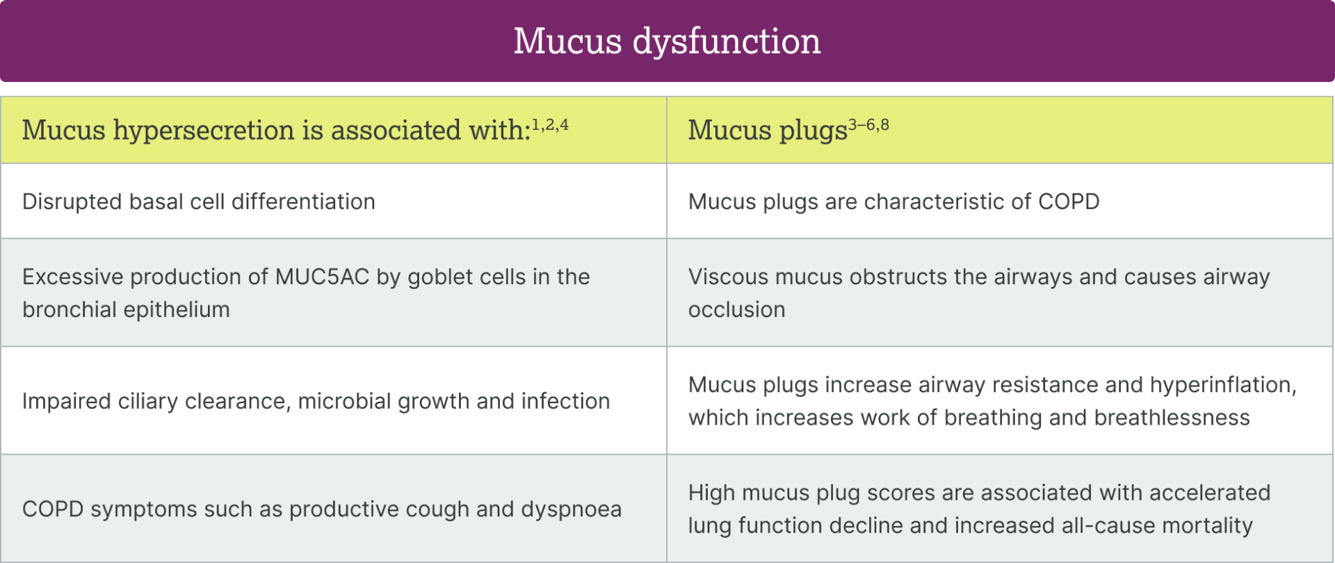
Mucus dysfunction
in COPD

Explore the latest in COPD science
  • Home
  • Burden of COPD
  • Inflammation in COPD
  • Mucus dysfunction in COPD
  • IL-33 in COPD
  • Resources

Snapshot

Mucus dysfunction is central to
COPD pathogenesis1–5

Productive cough and excess sputum correlate with higher disease burden, while mucus plugs are associated with increased exacerbation risk, faster FEV1 decline and higher
all-cause mortality2,3,6

There is poor correlation between mucus hypersecretion and mucus plugs, suggesting these processes may be partially independent and involve complex, overlapping mechanisms4,5,7

Mucus dysfunction is central to
pathology
in COPD1–5,8

View the data

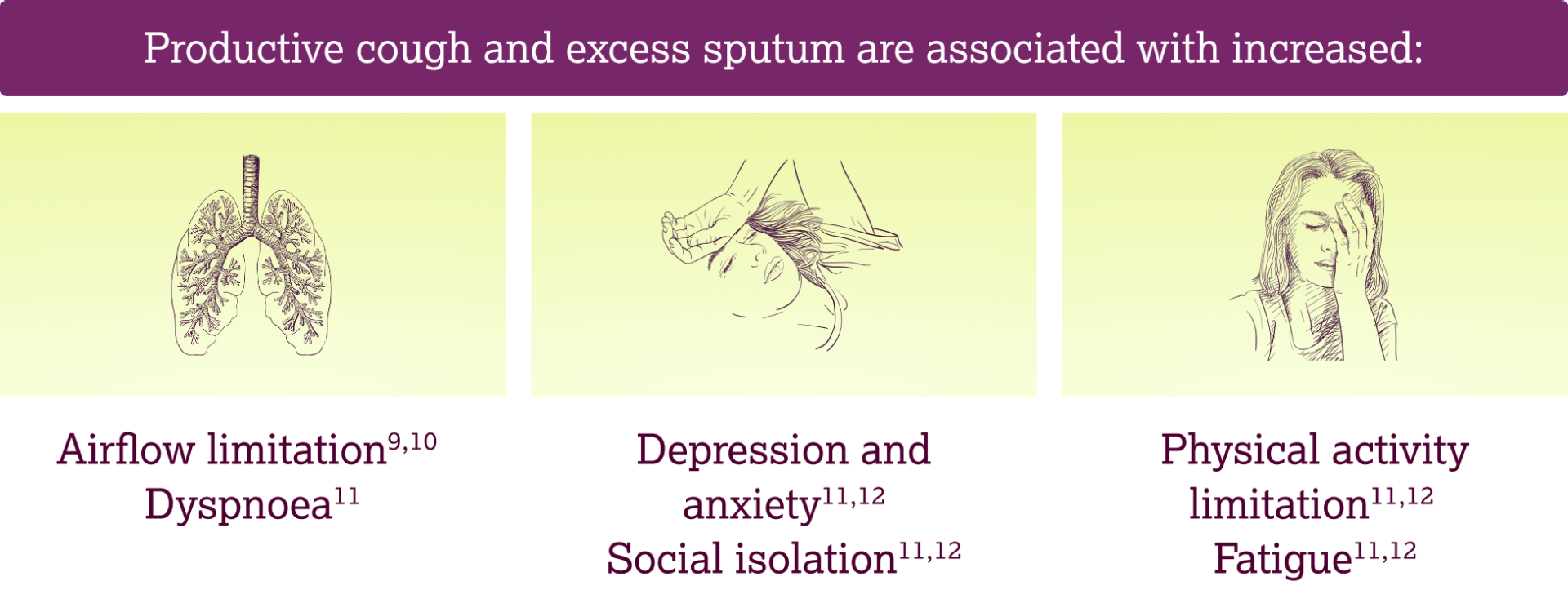
Mucus hypersecretion is associated with high disease burden3,9–12

View the data

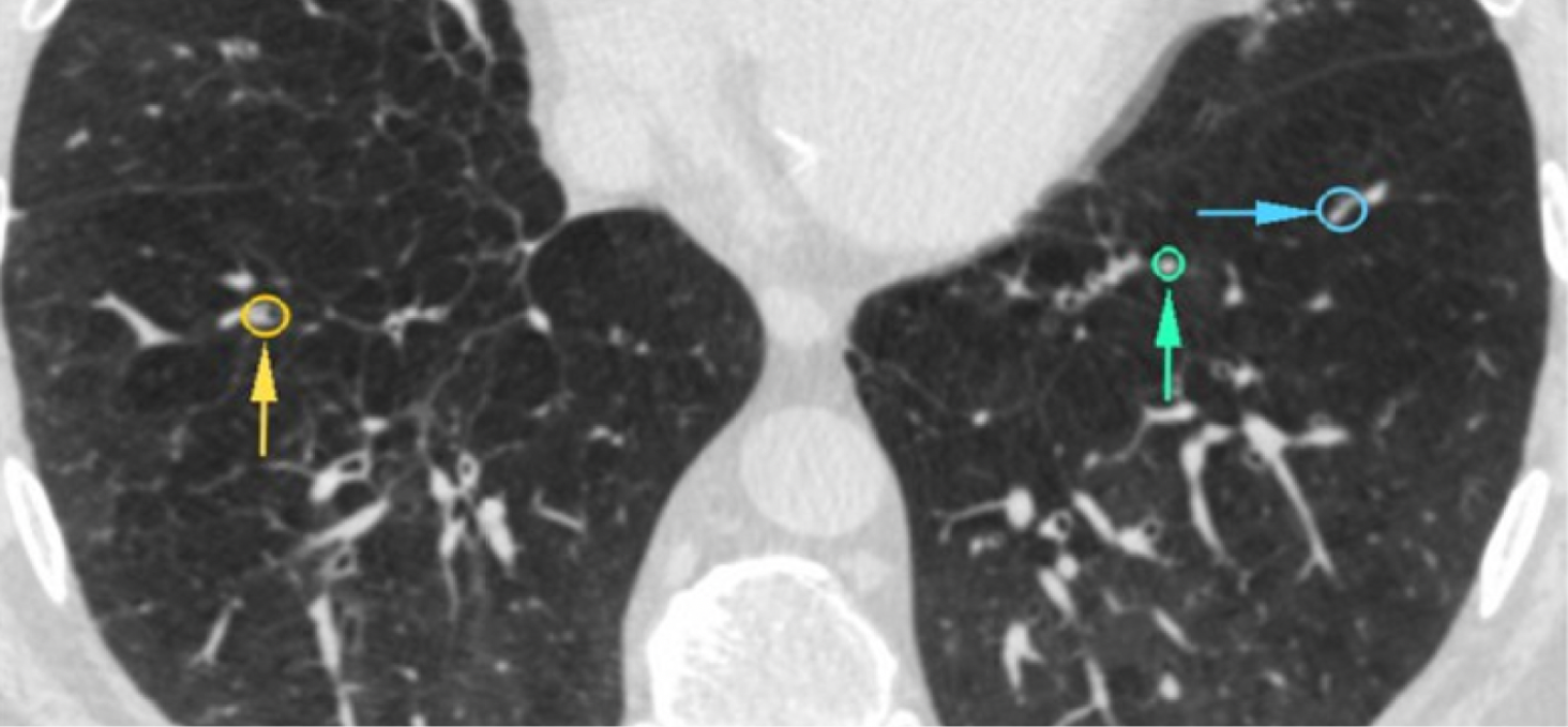
Mucus plugs occluding the airways have been observed on CT scans in 25–67% of patients with COPD3,5,13

View the data

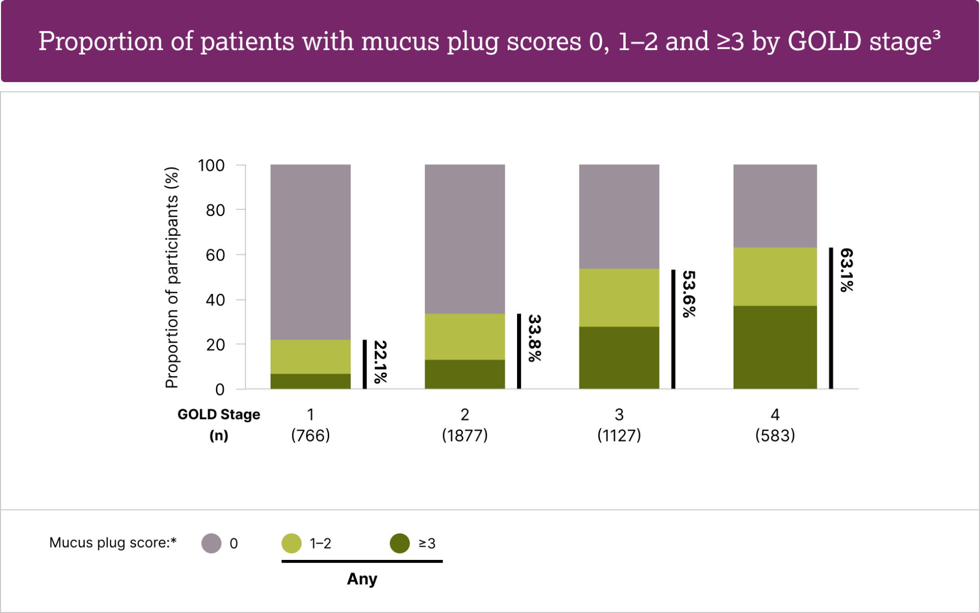
Mucus plugs increase in prevalence
with
disease severity3

View the data

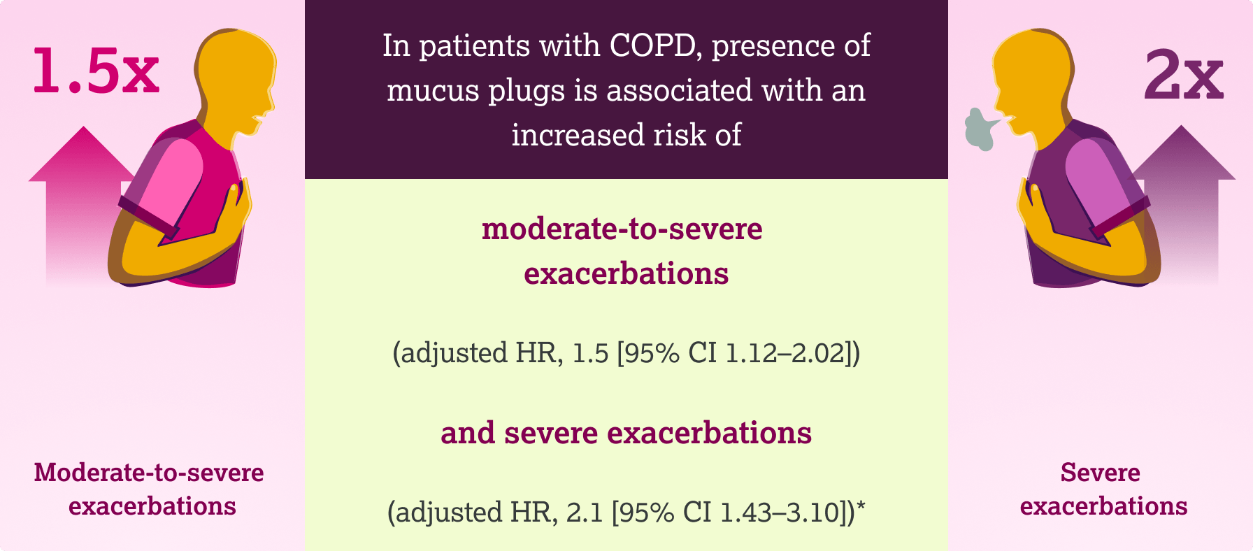
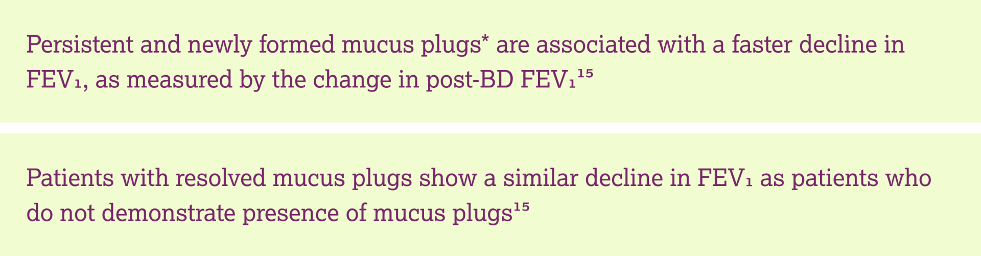
Mucus plugs are associated with increased risk of exacerbations6, increased risk of all-cause mortality3 and lung function decline15

View the data

There is a poor correlation between mucus hypersecretion and
mucus plugs7

View the data
  1. <h4>Over half of patients with COPD receiving triple inhaled therapy have been shown to continue to experience exacerbations<sup>1,2</sup></h4>
  2. <h4>Over half of patients with COPD receiving triple inhaled therapy have been shown to continue to experience exacerbations<sup>1,2</sup></h4>
  3. <h4>Over half of patients with COPD receiving triple inhaled therapy have been shown to continue to experience exacerbations<sup>1,2</sup></h4>
  4. <h4>Over half of patients with COPD receiving triple inhaled therapy have been shown to continue to experience exacerbations<sup>1,2</sup></h4>
  5. <h4>Over half of patients with COPD receiving triple inhaled therapy have been shown to continue to experience exacerbations<sup>1,2</sup></h4>
  6. <h4>Over half of patients with COPD receiving triple inhaled therapy have been shown to continue to experience exacerbations<sup>1,2</sup></h4>

Inflammation in COPD

Previous

IL-33 in COPD

Next

Frequently asked questions

What does ‘dysregulated IL-33’ mean?

IL‑33 is released upon stress or cell damage to initiate a host response—what was adaptive turns maladaptive or ‘dysregulated’ when its levels chronically surpass buffering mechanisms and signalling is persistent1–8

How does smoking affect the IL-33 pathway in patients with COPD?

In COPD, smoking status affects the IL‑33 axis—former smokers show elevated IL-33 compared to current smokers, while current smokers show reduced sST2 and more oxidized IL‑33, indicating aberrant IL‑33–ST2 signalling in both groups9–12

How does the role of IL-33 in COPD differ from other epithelial cytokines?

Damage to the epithelium triggers the release of upstream, alarmin cytokines. IL-33 is pre-formed, rapidly released and initiates a broad inflammatory cascade encompassing Type 1, Type 2 and Type 3 responses1,13,14

What evidence would suggest my patient with COPD has dysregulated IL-33 signalling?

Frequent exacerbations, productive cough and persistent airway/systemic inflammation may suggest heightened IL‑33

pathway activity15–17

What are the potential consequences of dysregulated IL-33 signalling?

IL-33 through its dual forms can drive Type 1 and 3 and Type 2 (eosinophilic, subset) inflammation, and mucus dysfunction, which are key drivers of COPD pathogenesis1–3,12,15

What is mucus dysfunction and what is the impact for patients with COPD?

Mucus dysfunction is the pathological changes in mucus volume, composition and clearance, the airway manifestations of these pathological changes, and the downstream

clinical consequences12,15,18–25

  1. sss
  2. sss
  3. sss
  4. sss
  5. sss
  6. sss

  1. Cayrol C. Cells. 2021;11(1):107
  2. Calderon AA, et al. Eur Respir Rev. 2023;32(167):220144
  3. Brightling CE, et al. Eur Respir J. 2019;54(6):1900651
  4. England E, et al. Sci Rep. 2023;13(1):9825
  5. Rabe KF, et al. Am J Respir Crit Care Med. 2023;208(4):395–405
  6. Keddache S, et al. Clin Immunol. 2021;229:108798
  7. Scott IC, et al. Biochem Soc Trans. 2018;46(5):1345–1353
  8. Burgoyne RA, et al. Cells. 2021;10(10):2763
  9. Abdo M, et al. Eur Respir J. 2024;64(3):2400347
  10. Faiz A, et al. Am J Respir Crit Care Med. 2023;208(10):1075–1087
  11. Faiz A, et al. Am J Respir Crit Care Med. 2023;208(Suppl.):1075–1087
  12. Strickson S, et al. Eur Respir J. 2023;62(3):2202210
  13. Cayrol C, Girard JP. Cytokine. 2022;156:155891
  14. Varricchi G, et al. Eur Respir J. 2024;63(4):2301619
  15. Diaz AA, et al. JAMA. 2023;329(21):1832–9
  16. Shen Y, et al. Int J Chron Obstruct Pulmon Dis. 2018;13:399–407
  17. Rodrigues SO, et al. Pharmaceuticals (Basel). 2021;14(10):979
  18. Fahy JV, Dickey BF. N Engl J Med. 2010;363(23):2233–2247
  19. Stott-Miller M, et al. Int J Chron Obstruct Pulmon Dis. 2020;15:2467–2476
  20. Hughes R, et al. Respir Med. 2022;200:106921
  21. Choate R, et al. Chronic Obstr Pulm Dis. 2020;7(1):49–59
  22. Cook N, et al. Int J Chron Obstruct Pulmon Dis. 2019;14:1365–1376
  23. Tian PW, Wen FQ. J Transl Int Med. 2015;3(3):89-92
  24. Mettler SK, et al. N Engl J Med. 2025;392(19):1973–1975
  25. Zhou D, et al. Expert Rev Respir Med. 2025:1–7

Stay ahead

Sign up for the latest updates on new content

Sign up

Back to top

BD, bronchodilator; CI, confidence interval; COPD, chronic obstructive pulmonary disease; CT, computed tomography; FEV1, forced expiratory volume in one second; GOLD, Global Initiative for Chronic Pulmonary Obstructive Disease; HR, hazard ratio; MUC5AC, mucin-5 AC

  1. Raby KL, et al. Front Immunol. 2023;14:1201658
  2. Kotlyarov S. Int J Mol Sci. 2022;23:985
  3. Diaz AA, et al. JAMA. 2023;329:1832–1839
  4. Fahy JV, Dickey BF. N Engl J Med. 2010;363:2233–2247
  5. Okajima Y, et al. Chest. 2020;158:121–130
  6. Jin KN, et al. Arch Bronconeumol. 2025;61:138–146
  7. Mettler SK, et al. Chest. 2024;166:1010–1019
  8. Gagnon P, et al. Int J Chron Obstruct Pulmon Dis. 2014;9:187–201
  9. Stott-Miller M, et al. Int J Chron Obstruct Pulmon Dis. 2020;15:2467–2476
  10. Hughes R, et al. Respir Med. 2022;200:106921
  11. Choate R, et al. Chronic Obstr Pulm Dis. 2020;7:49–59
  12. Cook N, et al. Int J Chron Obstruct Pulmon Dis. 2019;14:1365–1376
  13. Dunican EM, et al. Am J Respir Crit Care Med. 2021;203:957–968
  14. Farooqui ON, et al. Am J Respir Crit Care Med. 2026;212(2):268–76
  15. Mettler SK, et al. N Engl J Med. 2025;392:1973–1975

mucus-m1 mucus-m1

mucus-mod2 mucus-mod2

mucus-mod3
Representative CT images of a patient with COPD who showed mucus plugs in the airway14*

*Computed tomographic lung scans from 31 patients with COPD from the SubPopulations and InteRmediate Outcomes In COPD Study (SPIROMICS) cohort.


Annotated computed tomography of mucus plugs in patients with chronic obstructive pulmonary disease, showing the heterogeneity of size and shape.


Coloured arrows point to an elliptical region showing the mucus plugs visible on the lung scans.


Reproduced from Farooqui ON, et al. Mucus plugs in chronic obstructive pulmonary disease are heterogeneous in size and shape and occur in relatively large airways. Am J Respir Crit Care Med. 2026;212(2):268–76. doi: 10.1093/ajrccm/aamaf138. Article distributed under the terms of the CC BY 4.0 license.

mucus-mod4 mucus-mod4

Mucus plug scores determined from CT scans from COPDGene study participants3

*Mucus plug score calculated from the number of lung segments with mucus plugs. Mucus plugs were defined as an opacity that completely occluded the lumen of an airway3

Figure developed using Table 1 from: Airway-Occluding Mucus Plugs and Mortality in Patients With Chronic Obstructive Pulmonary Disease by Diaz AA, et al. JAMA. 2023;329:1832–1839. https://jamanetwork.com/journals/jama/fullarticle/2805343

Increased risk of exacerbations6
mucus-m5 mucus-m5

*Results are from a retrospective observational study evaluating patients in Korea (n=623) who were regularly followed up for COPD management at a teaching hospital between January 2004 and December 20196

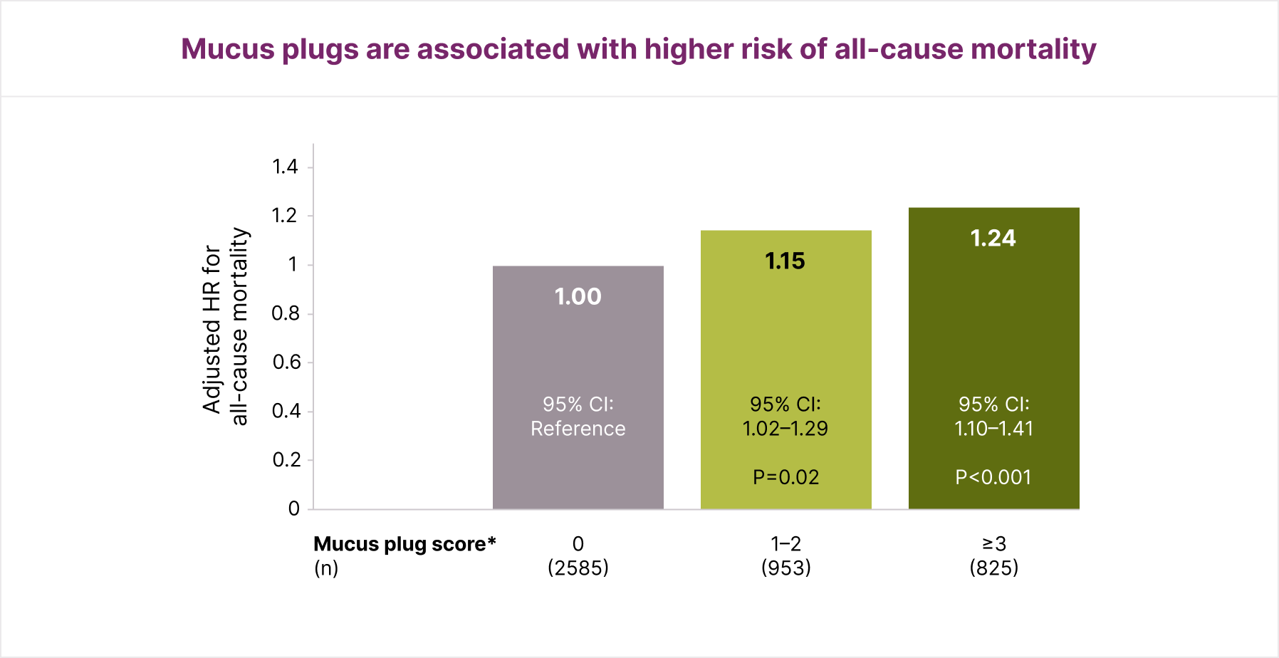
Increased risk of all-cause mortality3
mucus-m5b

Results are from an observational retrospective analysis of prospectively collected data from patients with a diagnosis of COPD, aged 45–80 years, with a smoking history of at least 10 pack-years. Participants were enrolled at 21 centres across the US between November 2007 and April 2011, and were followed up through August 31, 2022. 4363 patients were included in the primary analysis

*Mucus plug score assigned based on the number of lung segments with mucus plugs3

Figure developed using Table 2 from: Airway-Occluding Mucus Plugs and Mortality in Patients With Chronic Obstructive Pulmonary Disease by Diaz AA, et al. JAMA. 2023;329:1832–1839. https://jamanetwork.com/journals/jama/fullarticle/2805343

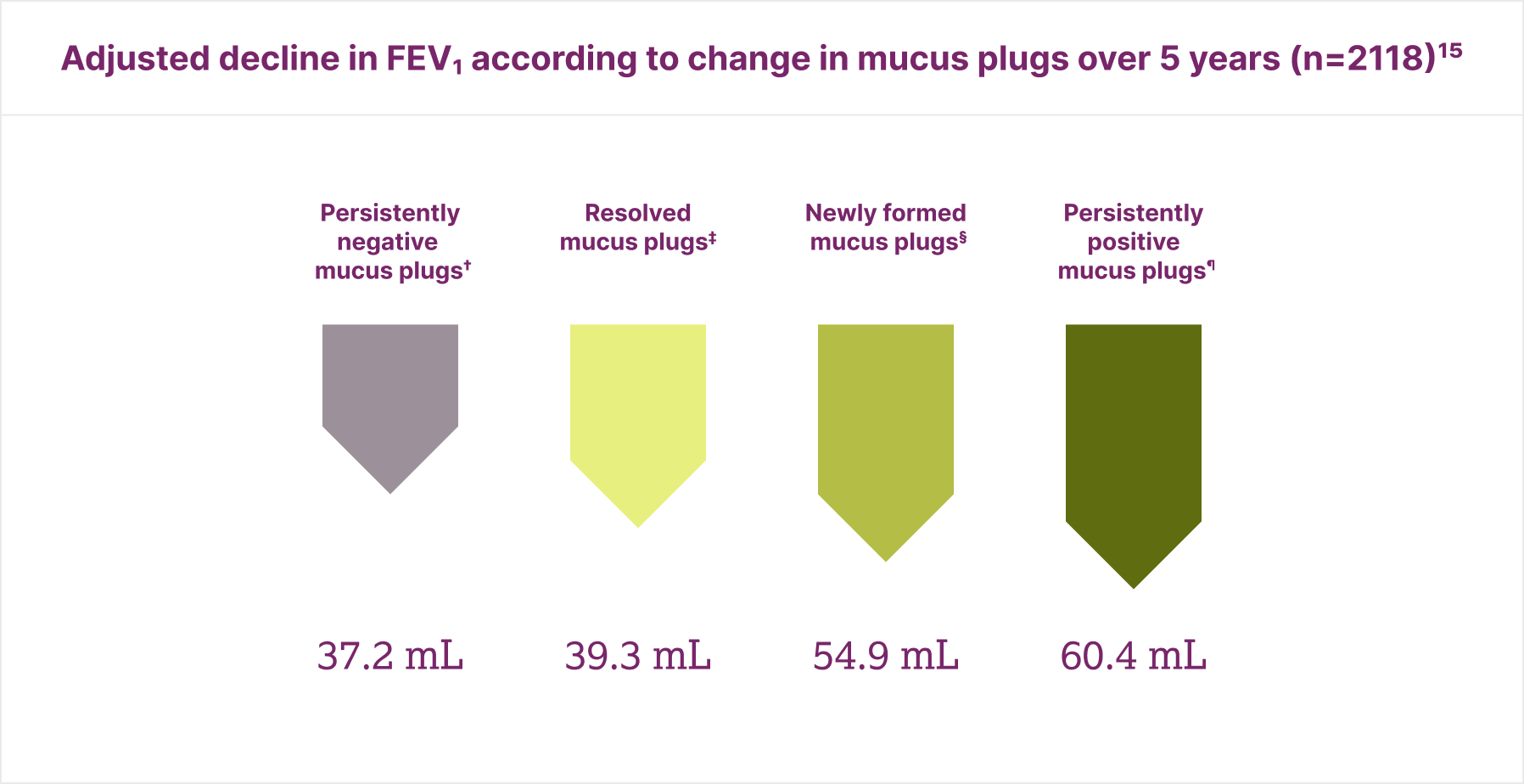
Lung function decline15
mucus-5c mucus-5c
mucus-5d

*Mucus plugs were surveyed with CT at baseline (phase 1) and at a 5-year follow-up visit (phase 2); †No mucus plugs observed at baseline or 5-year follow-up; ‡Mucus plug only present at baseline; §Mucus plug only present at 5-year follow-up; ¶Mucus plug present at baseline and 5-year
follow-up15

Figure developed using Figure 1B from: Longitudinal changes in airway mucus plugs and FEV1 by Mettler SK, et al. N Engl J Med 2025;392:
1973–1975. https://www.nejm.org/doi/full/10.1056/NEJMc2502456

mod6 mod6

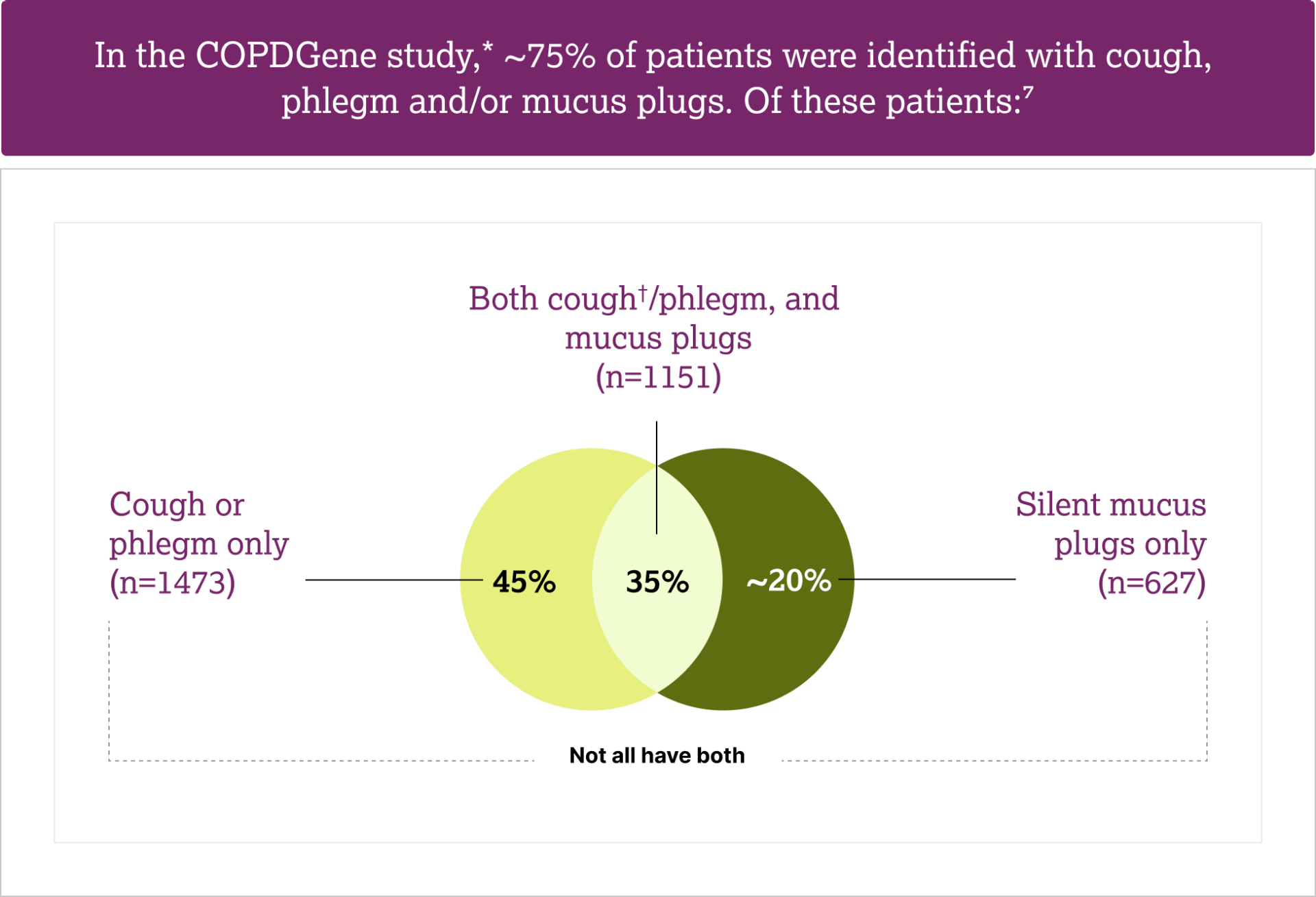
In the COPDGene study,* ~75% of patients (n=3251) were identified with cough, phlegm and/or mucus plugs and ~25% of patients (n=1112) had neither cough or phlegm, nor mucus plugs7


*Based on data from 4363 patients who previously smoked or with active tobacco use with the full spectrum of COPD severity. Patients were recruited from the COPDGene study, with available COPD and CT scan data15; †Included patients with both dry and productive cough7

Z4-86369 | Date of preparation: August 2026

Sign up below to be notified about new resource

Connessiallasalute.it

This site is intended for healthcare professionals


ASTRAZENECA Z4-85831 | 8/2026 |  ©ASTRAZENECA 2025 ALL RIGHTS RESERVED


Cookies

Contact Us

Privacy Notice